Ultrasonic

An ultrasound is a simple, painless, and safe examination, which is done using ultrasound (high frequency sound waves), without the use of radiation.

It gives us information about the functioning of the body's solid organs and vessels.

The accuracy and quality of the examination depends on:

  • The experience of the examiner (operator-dependent) is very important in the diagnosis. The diagnosis is made at the same time as the examination and the images are those taken by the examiner during the examination depending on the diagnosis;
  • The use of high-resolution multi-crystal machines.
Υπέρηχοι

EQUIPMENT

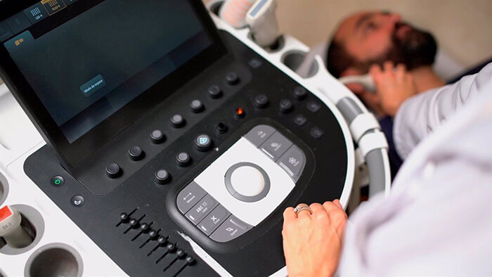
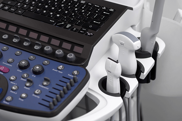
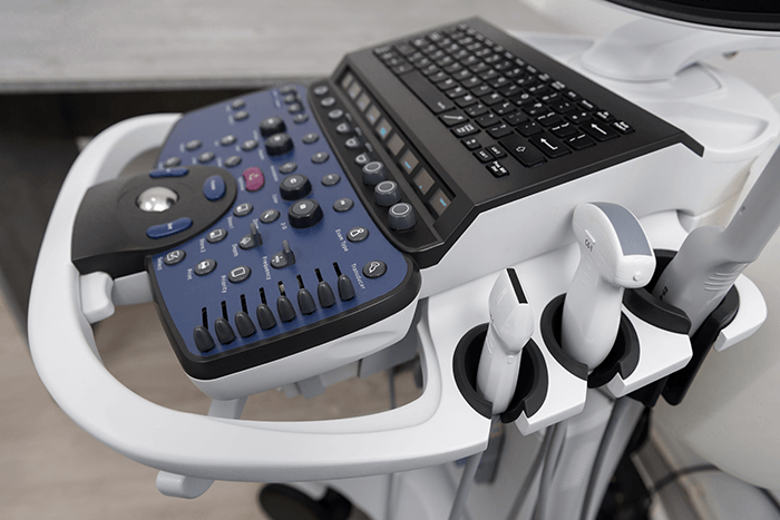
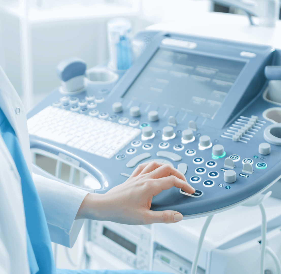
The ultrasound department is equipped with specialized medical staff and modern ultrasound machines.

PHIPIPS AFFINITY 70G KAI GE LOGIQS7 EXPERT, with high resolution sounders, tissue blood flow quantification system, with elastography technique.

Specialized examinations and invasive procedures are carried out with the standard of international centers abroad.

SPECIAL EXAMINATIONS

In addition to routine examinations, ultrasounds of organs and color Doppler (Triplex) of vessels, the following examinations are carried out as well:

  • • Study of solid organs (liver, kidney, spleen, pancreas);
  • • Preoperative vein mapping;
  • • Elastography of organs (thyroid, breast, liver, prostate, and other organs).

INVASIVE EXAMINATIONS

  • Rectal ultrasonography of the prostate using elastography and guided prostate biopsy
  • Fine Needle Aspiration (FNA) of thyroid - salivary glands - lymph nodes
  • Thoracic puncture (diagnostic - evacuation)
  • Ascitic collection puncture (diagnostic - evacuation)
  • Guided histological liver biopsy
  • Drainage of intra-abdominal abscesses - collections

DIAGNOSTIC EXAMINATIONS - ULTRASOUND OF ORGANS

  • UPPER ABDOMEN
    (Liver - biliary - pancreas - spleen).
    Preparation: The patient must not eat for 6 hours before the examination.
  • OWER ABDOMEN (SUPRAPUBIC AND INTRAVAGINAL)
    MALE: KIDNEYS, BLADDER, PRE/POST URINATION, PROSTATE
    WOMEN: KIDNEYS, BLADDER, GENITALS (UTERUS - OVARIES)
    Preparation: After urinating, the patient should drink 3-4 glasses of water 2 hours before the examination and not urinate until the examination is done.
  • THYROID ULTRASOUNDwith a color Doppler ultrasound study
  • THYROID ELASTOGRAPHY
  • CERVICAL ULTRASOUND with a color Doppler ultrasound study
  • CERVICAL ELASTOGRAPHY
  • ULTRASONOGRAPHY OF THE PARATHYROIDS
  • DYNAMIC MUSCOSKELETAL ULTRASOUND (Shoulder, Elbow, Hip, Knee, Muscle)
  • CAROTID & VERTEBRAL ARTERIES
    Color Doppler (Triplex) of carotid & vertebral arteries (hemodynamic and morphological study).
  • TEMPORAL ARTERIES
    Color Doppler (Triplex) of temporal arteries (hemodynamic and morphological study).
  • JUGULAR VEINS
    Color Doppler (Triplex) of jugular veins (hemodynamic and morphological study).
  • ABDOMINAL AORTA
    Color Doppler (Triplex) of abdominal aorta (hemodynamic and morphological study).
  • RENAL ARTERIES
    Color Doppler (Triplex) of renal arteries (hemodynamic and morphological study).
  • RENAL VEINS
  • ULTRASOUND OF HIPS IN NEWBORNS
    It is the main examination for the early diagnosis of hip dysplasia. It takes place during the first trimester after the birth of the child. In multiple states it is performed as a screening test in all newborns Guided Breast Biopsy for cytological and histological examination